


When and where was Photo 51 taken?
The photo was taken in May 1952 by Rosalind Franklin and her PhD student Raymond Gosling in the basement underneath the chemistry laboratories at the MRC Biophysics Unit. Franklin, a biophysicist, had been recruited to the unit to work on the structure of DNA. The unit was then part of the King’s College campus on the Strand in London. It was run by Sir John Randall, who had turned some of the university’s physics department over to studying biological problems.
The MRC Biophysics Unit moved to Drury Lane in the 1960s and later became the Randall Institute. I now work in its most recent incarnation, the Randall Centre for Cell and Molecular Biophysics at King’s College’s Guy’s Hospital campus. So, Photo 51 is doubly significant for me. I’m an X-ray crystallographer so it’s part of my heritage in that respect, but all of us in the centre are proud of this link with the work in the 1950s.
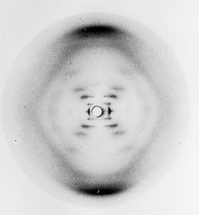
Photo 51. Credit: King’s College London
What is X-ray crystallography?
It’s a long-established method of determining the structure of molecules by bombarding them with X-rays. The molecules are in a crystal or otherwise ordered form, so when the X-rays bounce off the electrons in the molecule’s atoms, they scatter in a particular unique pattern. You can use that pattern to infer the structure. These days we take thousands of images from different angles and digitally build up a 3D image of the structure.
How would it have been done in the 1950s?
The technique in principle wouldn’t actually have differed too much, although it would have been a much more painstaking and time-consuming process. Franklin and Gosling used a very pure form of DNA and they became expert in pulling it into strands for analysis. Within each strand would have been a vast number of DNA helices lined up next to each other.
The DNA strand was fixed to a support and sealed in a camera, in front of a piece of X-ray film, and then exposed to X-rays for days at a time. Rather dangerously, hydrogen was bubbled through water and into the camera to stop the X-rays from bouncing off molecules in the air.
The film was then developed and the patterns emerged before the researchers’ eyes. Raymond Gosling often spoke of the great excitement of developing the films in the King’s basement.
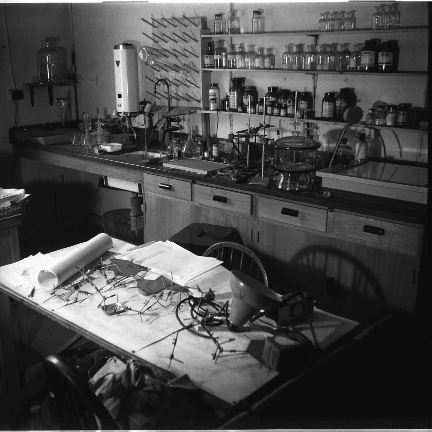
Rosalind Franklin’s lab at Birkbeck College. Credit: @JohnFinch/MRC Laboratory of Molecular Biology
What are we actually looking at when we look at Photo 51?
Photo 51 is an image of the more hydrated ‘B’ form of DNA. Franklin and Gosling had been experimenting with whether the humidity at which they kept the samples would affect the images. They had taken a series of images, and Photo 51 was taken at the highest humidity, around 92%.
The darker patches indicate where the film has been repeatedly bombarded by diffracted X-rays from regular, repeating features within the molecule. The dark patches at the top and bottom of the picture, for example, represent DNA’s ‘bases’, the four parts of DNA which make up the genetic code. These patches are dark because there are so many bases all arranged in a regular fashion.
You can work out the distance between bases in the structure by measuring the distance between the dark patches on the film. This involves a calculation based on how far the DNA sample was from the X-ray film and how it was orientated in the X-ray beam.
What about the cross shape of spots?
For people like Watson and Crick, who were already building models, this cross really spells out helix. Maurice Wilkins, who had worked on DNA separately from Franklin, showed this photo to Jim Watson when he came to visit and it really excited him.
A lot has been said and written about that moment and some people think that Wilkins shouldn’t have shared the photo, certainly not without Franklin’s knowledge or permission. But he had it legitimately as part of Rosalind’s papers as she was soon to leave for Birkbeck College. He was keen that research on the structure progressed, particularly because he wanted the UK to beat Linus Pauling in the US to discovering the structure.
The reason that the cross indicates a helix is that the arms of the cross represent the planes of symmetry in a helix viewed from the side. The ‘zig’ and the ‘zag’, so to speak, of the turns of the helix. It’s difficult to see clearly, but there are ten blobs on each arm of the cross before you reach the large black patch at the top.
This tells you that there are ten bases stacked one on top of the other in each turn of the helix. In fact, one of the blobs is missing, the fourth if you count out from the centre of the pattern. This indicates that one strand of DNA is slightly offset against the other.
If Franklin had all this information, why didn’t she suggest the structure?
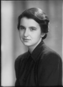
Rosalind Franklin by Elliott and Fry. Credit: © National Portrait Gallery, London
Well, it’s difficult to say but one reason is probably that Rosalind had chosen to focus her attention on her X-ray photos of a less hydrated ‘A’ form of DNA. This form appeared to show much more information and she hoped to calculate the structure directly, rather than build models. In fact, these photos of the ‘A’ form had revealed a key piece of information, namely that the two strands of DNA ran in opposite directions. Neither Rosalind nor the others had appreciated this, until Francis Crick realised its significance just before building the final model.
She didn’t turn her attention to Photo 51 until early in 1953. You can see from her notebooks that once she did concentrate on it, she gleaned all the key information about the structure from it. I fully believe that given more time she would have cracked the structure. She was so close. Watson was surprised that she accepted the correctness of their model immediately upon seeing it. It must have been because she could see that it fitted so well with all of her evidence.
What happened after the structure was published?
Franklin was already working at Birkbeck College by the time Franklin and Gosling’s paper, showing Photo 51, was published in Nature, alongside that of Watson and Crick’s model. Of course, Watson and Crick’s model was just that, only a model, so it needed to be verified. Wilkins built the first accurate model of DNA in the summer of 1953 and checked it against diffraction data such as Photo 51. Of course, the structure was right, it was too beautiful not to be.
This blog post has been refreshed from our archive.
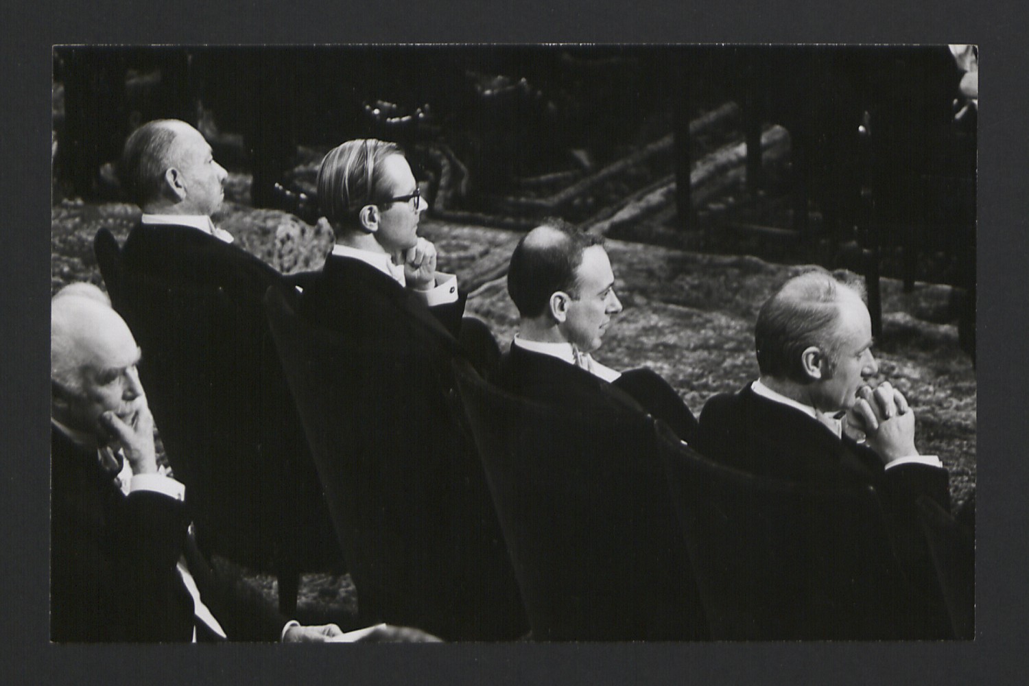
Maurice Wilkins, James Watson and Francis Crick at the 1962 Nobel Prize ceremony (K/PP178/15/3/1). Credit: King’s College London

Brian’s research over many years at King’s has involved the application of X-ray crystallography to study the molecular structures of antibodies, in particular those involved in allergy and asthma.
He was a founding member of the MRC-Asthma UK Centre for Allergic Mechanisms of Asthma. His work has led to the development of potential new therapeutics for allergic disease.