When Andrew first saw scientists growing tiny versions of his own eye cells in a laboratory dish, he gave them a nickname: “mini mes”.
These tiny clusters of cells, grown from a small skin sample taken from Andrew’s arm, are helping researchers investigate Best disease, a rare inherited eye condition that has affected his family across generations.
“It was brilliant,” Andrew says of learning his donated cells had become part of the research. “Whether it will do anything for me, I don’t know, but I’m hoping that at some point there may be an opportunity to correct the vision in our daughter.”
Scientists at the London Project to Cure Blindness are using cutting-edge gene-editing technology to investigate the disease. They are combining stem cells, clustered regularly interspaced short palindromic repeats (CRISPR) gene editing and patient-donated cells to understand how the disease develops and how it might one day be treated.
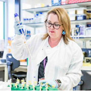
Associate Professor Amanda Carr working a lab. Credit: Ross Vickery
For Associate Professor Amanda Carr, who has spent more than 20 years researching inherited retinal diseases, that possibility marks a dramatic shift in what medicine can offer.
“When I first started working in inherited retinal disease,” she says, “there was really nothing we could do for patients. Now, these new technologies are opening doors we couldn’t even imagine before.”
Living with Best disease
Andrew has lived with Best disease all his life.
“I don’t consider it to be a disability because I don’t know anything different,” he says. “It’s always been part of my life.”
The condition affects central vision while leaving peripheral vision largely intact. That means he can get around independently, but everyday tasks such as reading, recognising faces and watching television can be difficult.
“If I look directly at your face, I can see your hair either side of it, but I can’t see any features,” he explains. “That’s why my eyes are always darting around, trying to pick up the detail.”
Unlike the black spot often used to illustrate sight loss, Andrew says that’s not quite how he experiences it.
“The blur takes on the colour of whatever I’m looking at. My brain fills in what’s missing.”
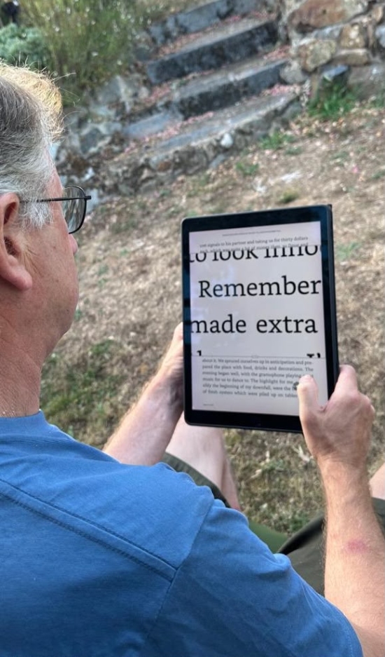
Andrew using a reading device to magnify text. Credit: Andrew
Modern technology has made a huge difference to his independence. “Phones and tablets have given me my independence back,” he says.
“I can photograph something and enlarge it, and I use the built-in magnifier and screen reader all the time. Lots of people don’t even realise those accessibility features are already there.”
Growing an eye disease in the lab
Best disease is caused by faults in a gene called BEST1, which affects a layer of cells at the back of the eye known as retinal pigment epithelial cells. These cells support the retina by supplying nutrients and clearing away waste. When they stop working properly, central vision gradually deteriorates.
To study the disease, Associate Professor Carr’s team starts with something surprisingly ordinary: a skin biopsy.
Researchers reprogramme skin cells into stem cells, then guide them into becoming retinal cells carrying exactly the same genetic mutation as the patient.
“It means we can watch what’s going wrong inside the cells in real time,” she says.
When the team grew Andrew’s retinal cells, they discovered an important protein called bestrophin wasn’t reaching the right place inside the cell.
“The cells looked swollen,” she explains. “It was like the chemical balance inside them had gone wrong.”
A different way to treat the disease
One of the biggest challenges is that there isn’t just one form of Best disease. Scientists have identified more than 500 different disease-causing mutations in the BEST1 gene.
Rather than designing hundreds of individual treatments, the team asked a different question: could they simply switch off the faulty copy of the gene while leaving the healthy copy untouched.
That idea became possible thanks to CRISPR gene editing.
Using naturally occurring genetic markers as signposts, the researchers identified the faulty copy of the gene in Andrew’s cells and selectively switched it off.
The results were striking.
The misplaced protein returned to the correct location, the cells began behaving more normally and harmful fatty deposits linked to the disease were dramatically reduced.
“It was really exciting,” Associate Professor Carr says. “We could see the cells recovering.”
Science with a human face
For her, meeting patients has transformed the atmosphere in the lab.
“A lot of science can feel abstract,” she says. “You’re working away with tubes and cells all day. Then you meet someone living with the disease and it completely changes how you think about the work.”
Andrew says the experience has been just as meaningful from the other side.